Thoracic trauma is the second most common cause of death in India following trauma (the most common cause is the head injury).
- Blunt trauma chest is the more common than penetrating trauma
- Ribs fracture is the most common type of chest trauma.
- Severe thoracic trauma dies during transport or after reaching the hospital, these deaths can be prevented by prompt diagnosis and intervention.
- Most chest trauma patients can be managed by Non-operative treatment.
- Only 10% of blunt trauma chest & 15-30% of penetrating chest trauma required Surgery.
- Physiological Triad of Thoracic Trauma – 1. Hypoxia, 2. Hypercarbia & 3. Acidosis.
- Most life-threatening thoracic trauma patients can be treated with – securing airway and chest decompression (Needle/ Finger or tube).
- Always remember – securing the airway is more important before chest decompression.
Thoracic Trauma – pattern
| Life-Threatening Thoracic Injury | Potentially Life-Threatening Thoracic Injury |
(Diagnose in Primary survey) | (Suspect in Primary Survey & diagnose in the secondary survey) |
| Tracheobronchial Tree Injury | Simple Pneumothorax |
| Tension Pneumothorax | Flail Chest |
| Open Pneumothorax | Hemothorax |
| Massive Hemothorax | Pulmonary contusion |
| Cardiac Tamponade | Blunt cardiac Injury |
| Circulatory cardiac Arrest | Traumatic Aortic disruption |
| Traumatic Diaphragmatic injury | |
| Blunt Esophageal Rupture |
Tracheobronchial Tree injury
- Case-A chest trauma patient with falling SpO2 – you intubated but SpO2 is not improving or even worsen (may be associated with Subcutaneous emphysema)- most probably you are dealing with TBT injury.
- Potential fatal injury and most difficult to diagnose – required expert to suspect injury & Prompt intervention to salvage the patient.
- Most common site – 1 inch (2.54cm) from the carina.
- Intubation can worsen the condition and kill faster.
- A high index of suspicion and prompt thoracotomy or fibreoptic intubation may save the patient.
Tension Pneumothorax
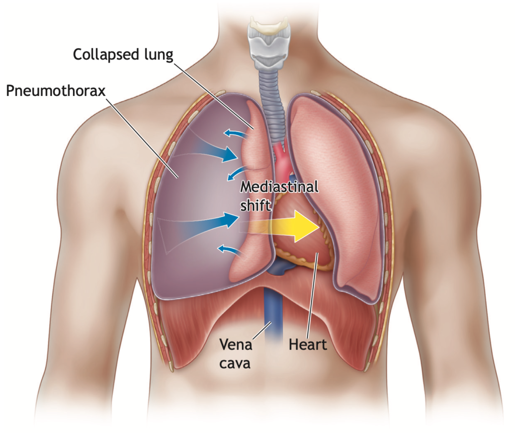
- Case – A chest trauma patient presented in your ED with breathlessness with Low O2 saturation, and hypotensive. Affected side absent breath sounds & no respiratory movement noted on that side – most probably you are dealing with Tension Pneumothorax, Now you have few seconds to save the patient.
- Pathophysiology – One-sided valve air leak
- Continuous expanding air shift mediastinum to the opposite side and leads to obstructive shock.
- Remember – Open pneumothorax can be converted into tension pneumothorax if the wound is sealed completely.
- Intervention – Immediate Needle/finger decompression followed by chest tube placement.
- Site – 5th Intercostal space at just anterior to the midaxillary line (According to 10th edition of ATLS). Few still prefer at 2nd ICS at the midclavicular line.
- Misinterpreting diagnosis with tension pneumothorax is cardiac tamponade.
- Attempting Needle decompression is not harmful as not attempting. (may kill your patient)
Open Pneumothorax
- Penetrating chest trauma
- Immediate 3 sided dressing followed by chest tube placement and closure of the wound.
- Never close the wound without putting a chest tube – that will kill your patient faster than not doing anything.
Massive Hemothorax
- 1500 ml of fresh blood comes immediately or 200 ml/ hr for 3-4 hours – leading to the patient being hemodynamically unstable.
- Immediately treated with ICD placement.
- Need urgent blood transfusion and thoracotomy to secure bleeding.
Cardiac Tamponade
- Most commonly associated with penetrating chest trauma
- Can be caused by Blunt trauma chest – usually after blunt cardiac rupture of the heart.
- Cardiac tamponade mimics – tension pneumothorax.
- In Tension Pneumothorax – air entry will be absent and in cardiac tamponade, air entry will be present.
- Beck’s Triad – 1. Hypotension, 2. Distended neck veins and 3. Distant (muffled) heart sounds.
- Intervention – Cardiocentesis follow by Thoracotomy
- CECT thorax is recommended in stable patients only.
- All Penetrating thoracic trauma cases leading to cardiac tamponade – Thoracotomy is mandatory.
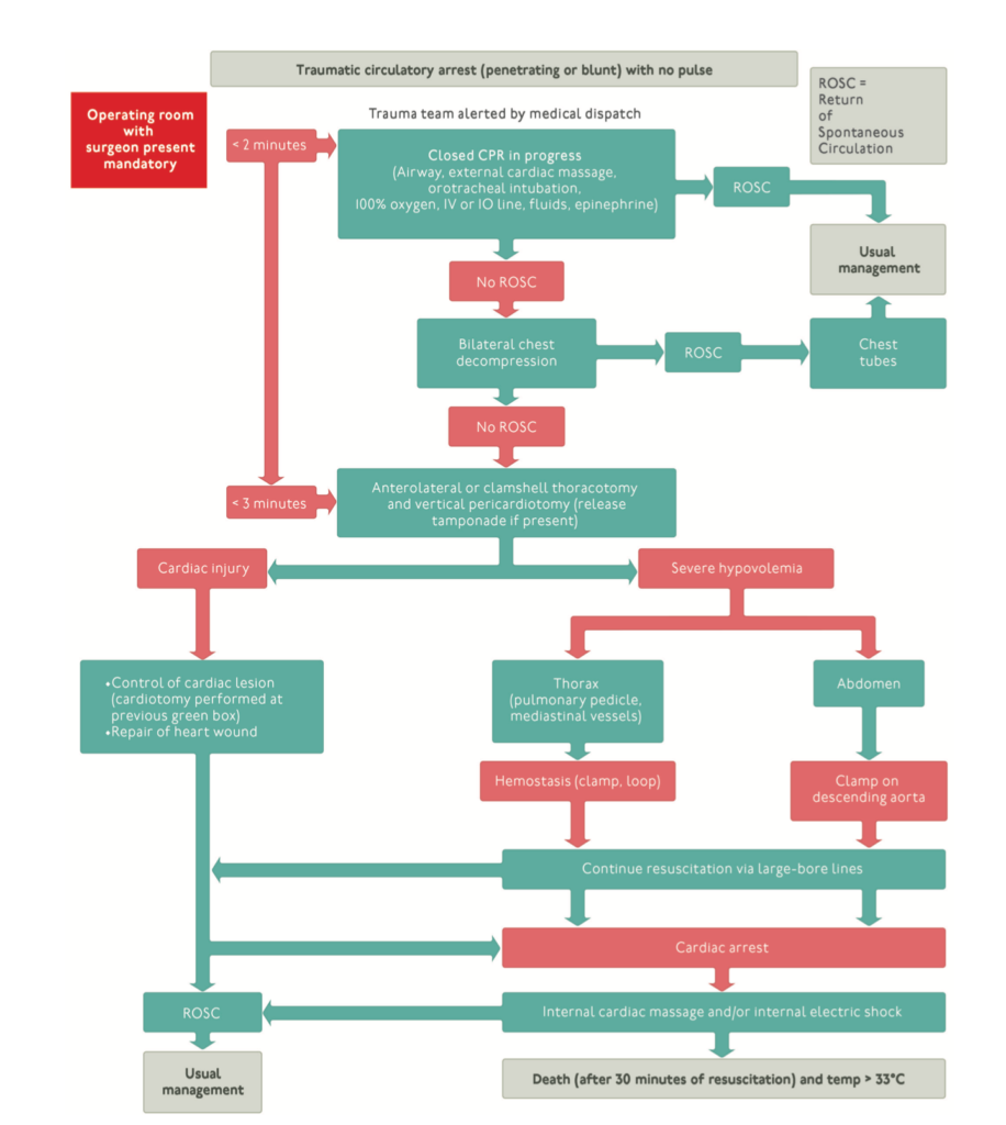
Traumatic cardiac arrest
Approach a patient with cardiac arrest patient following trauma – completely different from a non-trauma patient.

Simple Hemothorax or Pneumothorax
- Chest trauma is most commonly associated with multiple ribs fracture
- Simple pneumo or hemothorax is most commonly associated with Multiple ribs fracture.
- Diagnosed radiologically with chest x-rays or USG or CT
References
- Henry S. ATLS 10th edition offers new insights into managing trauma patients. Bulletin of the American College of Surgeons. 2018 Jun 1.
- Henry S. Earn CME.


Top ,.. top top … post! Keep the good work on !